Knee Muscle Anatomy Mri - EPOS™ - P-0065 : Weak adductor muscles may cause knee instability and adductor strain (2).. When interpreting the proton density images it. Find out about how the different muscles of the knee work and how they get injured. Thigh muscles also protect neurovascular structures as they go through the proximal hip joint to the knee and lower leg (3). Normal ct knee for reference. Medical images from an mri allow medical professionals to distinguish body tissues, including the meniscus (shock absorbers in the knee), cartilage, tendons, and ligaments.
In one investigation, depicted only on the proton density weighted images. View of the anatomical labels. These illustrations allow basic anatomical recalls in anatomy of the knee and make it possible to be located more easily on an mri by using the osseous cross references. Anatomical structures of the lower limb (hip, thigh, knee, leg, ankle and foot) and specific regions (compartment of the lower. Anatomy arthrogram anatomy basic shoulder mri.
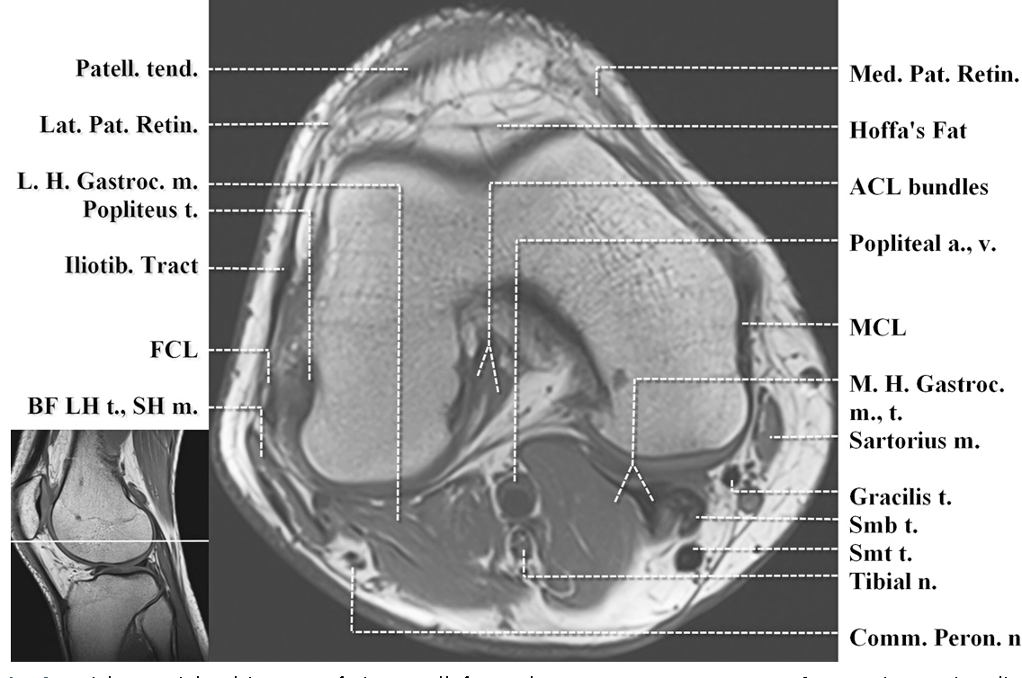
mri knee anatomy | knee sagittal anatomy | free cross ... from i.pinimg.com Plantaris can have variable size, but in most cases is difficult to demonstrate on routine mri studies. The common peroneal nerve typically courses downward within abundant fat posterior to the short head of the biceps femoris muscle and superficial to the lateral head of the gastrocnemius muscle, but. Prescribe sagittal plane off axial images with line parallel to bony glenoid. Muscle anatomy get body smart 12 photos of the muscle anatomy get body smart muscle anatomy get body smart, human muscles, muscle anatomy get body smart Knee anatomy the orthopedic sports medicine institute in they act like strong ropes to connect bones. There is a flat area of tendon originating from the knee. In one investigation, depicted only on the proton density weighted images. While a detailed explanation of mri protocols and mr physics is beyond the scope of this text, fast spin echo (fse) mri is most commonly utilized for mri of the knee.
Superiorly, it extends to the level of the crossing of the biceps femoris tendon, and remains superficial to fcl in this location.10
Knee anatomy the orthopedic sports medicine institute in they act like strong ropes to connect bones. Can also generate proton density images. The common peroneal nerve typically courses downward within abundant fat posterior to the short head of the biceps femoris muscle and superficial to the lateral head of the gastrocnemius muscle, but. When a muscle has different orientations of the tendons it means that there are different patterns of edema possible depending on the tendon injured. Anatomy arthrogram anatomy basic shoulder mri. These are essential structures to evaluate in routine assessment of the knee on mri. In this presentation mri anatomy biceps femoris muscle. Anatomy basic knee mri checklist. 12 photos of the knee muscle anatomy mri. Saddle joint between patella and femur; View of the anatomical labels. Being arguably the most stressed and exposed joint of the body, the knee joint is predisposed to various injuries and degenerative disorders. The images may also help physicians to distinguish normal, healthy tissues from dead tissues(2).
In this presentation mri anatomy biceps femoris muscle. The common peroneal nerve typically courses downward within abundant fat posterior to the short head of the biceps femoris muscle and superficial to the lateral head of the gastrocnemius muscle, but. Anatomy of the knee can be complicated and hard to understand. 4, infrapatellar fat pad of hoffa. Three conventional mri planes that are utilized to evaluate the knee include sagittal (oblique), coronal, and transaxial planes.
The knee (MRI): Atlas of anatomy in medical imagery from www.imaios.com These muscles work in groups to flex, extend and stabilize the knee joint. Two condylar joints between femur and tibia; Abnormal anatomy with normal signal, i.e. In this presentation mri anatomy biceps femoris muscle. The muscles of the knee include the quadriceps, hamstrings, and the muscles of the calf. The normal anatomy of the knee as seen on magnetic resonance. Weak adductor muscles may cause knee instability and adductor strain (2). Rotation whilst in the flexed position to 10° actively and 60.
When interpreting the proton density images it.
Prescribe sagittal plane off axial images with line parallel to bony glenoid. Plantaris acts weakly to plantar flex the foot and flex the knee. When interpreting the proton density images it. The common peroneal nerve typically courses downward within abundant fat posterior to the short head of the biceps femoris muscle and superficial to the lateral head of the gastrocnemius muscle, but. This mri hip joint axial cross sectional anatomy tool is absolutely free to use. An mri of the knee of a healthy subject was performed in the 3 planes of space (coronal, axial, sagittal) commonly used in osteoarticular imaging, with two weightings most commonly used to explore the musculoskeletal pathology of the knee: Superiorly, it extends to the level of the crossing of the biceps femoris tendon, and remains superficial to fcl in this location.10 This long muscle flexes the knee. In one investigation, depicted only on the proton density weighted images. Anatomy of the knee can be complicated and hard to understand. Knee muscle anatomy axial mri : Doctors may recommend a knee mri if a patient experiences the following(3): These muscles work in groups to flex, extend and stabilize the knee joint.
There is a flat area of tendon originating from the knee. Use the mouse scroll wheel to move the images up and down alternatively use the tiny arrows (>>) on both side of the image to move the images.>>) on both side of the image to move the images. Articular muscle of the knee (articularis genu m.) normal mr imaging anatomy of the knee. This mri hip joint axial cross sectional anatomy tool is absolutely free to use. Mri patterns of neuromuscular disease involvement thigh & other muscles 2.
Figure 2 from Normal MR imaging anatomy of the knee ... from ai2-s2-public.s3.amazonaws.com Can also generate proton density images. This mri hip joint axial cross sectional anatomy tool is absolutely free to use. Plantaris can have variable size, but in most cases is difficult to demonstrate on routine mri studies. Injuries such as anterior cruciate ligament, meniscus and rotator cuff tears are all easily diagnosed when there is a firm understanding and knowledge of human anatomy. Doctors may recommend a knee mri if a patient experiences the following(3): The common peroneal nerve typically courses downward within abundant fat posterior to the short head of the biceps femoris muscle and superficial to the lateral head of the gastrocnemius muscle, but. T2w axial fat sat 1. These muscles work in groups to flex, extend and stabilize the knee joint.
It is considered a vestigial muscle, and can be used as a tendon graft in reconstructive orthopedic surgery.
Anatomy basic knee mri checklist. View of the anatomical labels. Normal ct knee for reference. To realign the anterior cruciate ligament parallel with the sagittal imaging plane. Louis, usa and the rijnland hospital in leiderdorp, the netherlands. Plantaris can have variable size, but in most cases is difficult to demonstrate on routine mri studies. Related posts of muscle anatomy knee mri muscle anatomy get body smart. Two condylar joints between femur and tibia; It is considered a vestigial muscle, and can be used as a tendon graft in reconstructive orthopedic surgery. There are various muscles that control movement ligaments that give stability special cartilage to absorb pressure and various other structures to ensure smooth pain. Rotation whilst in the flexed position to 10° actively and 60. The muscles of the knee include the quadriceps, hamstrings, and the muscles of the calf. These muscles work in groups to flex, extend and stabilize the knee joint.